What is the difference between x-rays and fluorography, few know, however, many are interested. Relevant information is needed in order to understand what to do harmful and what not, and how often it is possible to undergo survey data. In addition to a different mechanism of influence, examinations are deciphered in different ways and used for various purposes.
Fluorography of the lungs is a special X-ray diagnostic technique, the essence of which is photographing the shadow of the organs of the chest itself, which is carried out using a fluorescent screen directly on the film. This method is still used despite the fact that it is very outdated. Today it is quite possible to translate it into a digital image.
But X-ray is a special study by fixing objects on film. It can be not only the lungs, but also all parts of the body.
X-ray of the lungs and fluorography have a significant difference. Patients should understand that it is fluorography that is considered safer, since it is less radioactive and does not affect a person so negatively. But her problem is that she has a lower resolution, which can affect the very quality of the result.
What is fluorography and what is worth knowing for yourself
Absolutely everyone, faced with a direction for fluorographic research. It is he who is made as a “legal” screening of lung diseases. And most interestingly, without it, the doctor will not sign a medical commission.
To do fluorography today is very popular - in our country a large influx of patients with tuberculosis and there is a need to prevent the spread of the problem.
It should be understood that a study once a year is not harmful, since a single dose does not exceed 0.015 mSv, while the preventive dose is 1 mSv. All this suggests that an overdose from a procedure such as fluorography can only be done if it is done about 1000 times in one year. It is worthwhile to understand that without the appointment of a doctor and his wishes, you do not need to chase the passage of this procedure yourself.
Today, there are several types of fluorography:

Unfortunately, in our hospitals and clinics, rooms where such procedures are performed have old equipment. A survey must be prescribed in the following cases:
- FLH for those who are visiting a medical institution for the first time;
- be sure to go through the procedure and those who live with a pregnant woman or in a family where there is a newborn baby;
- those who undergo a medical commission before going to the army or those who enter the contract service;
- HIV infected.
According to legal standards, it is enough to carry out the procedure no more than twice a year.
What you should know about lung x-rays and how harmful it is
Radiography is essentially an alternative to fluorography itself, but it has its own plus - a high resolution. It is interesting that x-rays can show shadows in the image up to 2 mm, which can not be said about fluorography, where you can observe shadows of only 5 mm.
Such a procedure as an X-ray is prescribed for bronchitis, pulmonary tuberculosis, pneumonia, cancer, and so on. By the way, fluorography is considered a preventive method. The mechanism of the procedure itself is quite simple: certain areas are illuminated when x-rays pass through them. When the patient undergoes this procedure, he is irradiated.
In medical institutions we see old devices, characterized in that they irradiate the patient many times more than necessary and possible for humans. On new equipment from lung x-rays, harm is not observed at all. But when it comes to treating acute pneumonia, doctors don’t go through private clinics or state-owned clinics to select new equipment, as it is necessary to make an urgent diagnosis as quickly as possible. Irradiation on the device should not exceed 0.6 mSv per year, if we talk about old equipment, then people can get 1.5 mSv on it.

The modern digital method of fluorography has a smaller radiation effect on the patient’s body, while at the same time, lung x-ray is a more informative way to determine lung pathologies, but less safe.
It should be understood that it is dangerous to do an x-ray in the following cases:
- During pregnancy;
- Before the planned conception.
With pneumonia, the doctor may prescribe an X-ray. To undergo such a procedure, the patient does not need to prepare in advance, and take additional items with him. There is the only condition that is required in order to make an x-ray correctly - to remove all unnecessary accessories from the chest (chains, laces and so on). It is not necessary to undress, you can stay underwear (but without iron fasteners).
There are two types of lung radiography:

The ultimate goal of the procedure is to get a special picture, examining which, the doctor can determine the diagnoses to prescribe a course of treatment. Of course, deciphering such a photograph is difficult. A specially trained person is engaged in this. He will easily study the forms of dimming and enlightenment, and he will also be able to consider the intensity of the lines and their shade and, through all the material, will be able to conclude about the work and pathology of internal organs. For example, lung cancer in the image will be depicted as rounded spots of different diameters, but with clear boundaries. If the boundaries are not clear, but blurred, then this will indicate cardiovascular disease or pneumonia. But tuberculosis in the image will be depicted as intense lines in combination with small, darkened areas.
Doses of radiation and whether it is possible to replace one method with another
X-ray or fluorography, which is better and what differences are they characterized by? In fact, these are two radiological examinations of the chest. But how do they differ? Of course, they are associated with radiation, and the dose of radiation itself depends not only on the research method, but also on the equipment itself and its characteristics.
Fluorography is usually done with only one image, which cannot be said about X-rays, which are done in several projections. If we talk about FLH, then the patient receives a dosage of 0.5 vZm, but with X-ray (in each of the two projections) - 0.5 vZm.
Fluorography and X-ray of the lungs what is the difference? In the first version, we get a very small picture. If we are talking about a small shot, then this is 30 * 30, and if about a large shot - 70 * 70. X-ray allows you to get a larger image, which will allow you to examine the organs in more detail.
It is logical that fluorography saves the film, since the picture is very small, but the normativity of the method is reduced, and this suggests that it is difficult to make an accurate diagnosis.

During pregnancy, breastfeeding, as well as planning, it is not necessary to conduct both x-ray and fluorographic studies of the organs of the chest cavity.
What is better x-ray or x-ray? Is it possible to replace one with another? X-ray is in its essence the simplest and most informative method for studying human internal organs and bones. But the fluorographic examination is simply aimed at identifying lung diseases. The principle lies in both cases, one and the same, but for all that - the goals are different. Say whether it is possible to do one thing instead of the other incorrectly.
- the radiation dose is not so high;
- ease and simplicity in passing, minimal waste of time;
- can determine the problem in the patient, after which it will be possible to refer for further examination and treatment.
It should be understood that no one prescribes X-rays as screening, therefore, there is the superiority of fluorography.
Also, many are interested in the question, can I do x-rays after fluorography? When a person goes to fluorography and receives unsatisfactory results, he can be sent for x-ray. But to do fluorography after x-ray is not recommended. If a person made an X-ray of the lungs, then it is logical that he does not need FLG. If he did an X-ray of the spine (where there is a large dose of radiation), there is no need to do fluorography immediately. It’s worth the wait.
Most often, the doctor prescribes an x-ray after fluorography in the following cases:
- if there is wheezing in the lungs;
- if the patient feels chest pain;
- if the patient has severe shortness of breath;
- with a strong and prolonged cough.
An X-ray of the lungs can diagnose the following diseases to the patient:

Is it possible to replace one with another? The question is serious. It is often believed that fluorography is harmful, but no x-ray, or vice versa. But in any case, a person must do something to clarify problems with the respiratory system. If you choose x-rays yourself, then all responsibility falls on the shoulders of the patient himself.
Speaking about the number of procedures performed, one can see the following: an X-ray of the lungs, the patient can do just as many times as the doctor tells him. If we talk about preventive measures, then the dose should not exceed 1 mSv per year. It is logical that, when prescribing, the physician must take into account the harm of the x-ray itself.
Where can I do x-rays or fluorography?
Since this procedure is related to irradiation, in order to get a detailed result, it is necessary that the equipment be very high quality and reliable. The older the equipment, the more likely it is to get an excess of radiation exposure and a poor-quality image. New devices give excellent picture quality and a minimum of harm to health. But it is almost impossible to find such equipment in a municipal institution, since there is mainly obsolete equipment there. In addition, in private clinics, undergoing fluorography on good equipment can be paid.
X-ray and fluorography are two ways of examining the internal organs of a person, without which most, diagnosis cannot be made. Treat this method of diagnosis carefully and not violate the regimen and recommendations for its passage. If you have undergone fluorography, you do not need to do it every three months, or four. It is enough to do 1 per year. And if you did FLG once, but lost its result, in the reference hospital you can take a copy so as not to do it again. If there is no need to make a regent, then it is logical not to do it just like that. Irradiation in large doses can be harmful, so be careful and attentive to these two studies.
The principle of operation of radiography and fluorography has a similar character, but these procedures are done on various equipment. But a person must undergo a fluorogram every year, and an x-ray is done only as prescribed by the doctor.
Consider the main distinguishing features of these diagnostics. In medical practice, an X-ray of the lungs differs from fluorography in that the doctor needs his result to make a diagnosis. Fluorography is performed to examine the organs of the chest and mammary glands.
During the radiography, the doctor receives the result on film. Therefore, this diagnostic method is recommended to be used in order to confirm or exclude the diagnosis. For example, pulmonary emphysema on an x-ray looks like an illuminated spot. And the larger it is, the greater the severity of the course of the disease.
Fluorography is mainly intended for making diagnoses such as tuberculosis, oncology, pneumonia. This method is the most superficial, and emphysema on the fluorogram is not indicated, even if the disease is started. Therefore, if there is a suspicion of any lung disease, it is better to conduct an x-ray examination.
The main part of the radiological equipment is a tube in which there is radiation. Rays penetrate the organ to be examined. Unlike x-rays, fluorography has a lower share of radiation, it is recommended for people from 14 years old to pass no more than once a year for the prevention of chest diseases. Both research methods are contraindicated in pregnant and lactating women.
During fluorography, a shadow picture is taken on a small format film from an optical and X-ray screen. Unlike film from radiography, the screen is highly sensitive. To obtain a small-format image, an X-ray fluorography apparatus is used. It is a fluorograph - a cabin with a radiation source. This type of diagnosis is used to detect oncology, tuberculosis, diseases of the lungs and bronchi.
Both radiography and fluorography have a negative effect on the human body. For X-rays, the radiation fraction is 1.5 m3v / g. With fluorography, this indicator is slightly lower. When conducting x-rays, the rays pass through the organ under study and the data is reflected on the film. This method is more accurate. On the fluorogram, the image is first visible on the screen, and then it is recorded on the camera.
Fluorography and radiography are prescribed depending on the symptoms and severity of the disease. There is also a radio photography method. It is used to detect tuberculosis and lung examination. For this technique, a stationary mobile device is used.
At the moment, in medicine, digital diagnostics are increasingly being used instead of film diagnostics. This study is much faster and easier. The picture is not saved on film, but in the database. This type of study is considered the safest for the human body.
Warning /var/www/x-raydoctor..php on line 1364
Warning: preg_match (): Compilation failed: invalid range in character class at offset 4 in /var/www/x-raydoctor..php on line 1364
Warning: preg_match (): Compilation failed: invalid range in character class at offset 4 in /var/www/x-raydoctor..php on line 1364
Warning: preg_match (): Compilation failed: invalid range in character class at offset 4 in /var/www/x-raydoctor..php on line 1364
Warning: preg_match (): Compilation failed: invalid range in character class at offset 4 in /var/www/x-raydoctor..php on line 1364
Warning: preg_match (): Compilation failed: invalid range in character class at offset 4 in /var/www/x-raydoctor..php on line 1364
Warning: preg_match (): Compilation failed: invalid range in character class at offset 4 in /var/www/x-raydoctor..php on line 1364
Warning: preg_match (): Compilation failed: invalid range in character class at offset 4 in /var/www/x-raydoctor..php on line 1364
Warning: preg_match (): Compilation failed: invalid range in character class at offset 4 in /var/www/x-raydoctor..php on line 1364
Warning: preg_match (): Compilation failed: invalid range in character class at offset 4 in /var/www/x-raydoctor..php on line 1364
Warning: preg_match (): Compilation failed: invalid range in character class at offset 4 in /var/www/x-raydoctor..php on line 1364
Warning: preg_match (): Compilation failed: invalid range in character class at offset 4 in /var/www/x-raydoctor..php on line 1364
Warning: preg_match (): Compilation failed: invalid range in character class at offset 4 in /var/www/x-raydoctor..php on line 1364
Warning: preg_match (): Compilation failed: invalid range in character class at offset 4 in /var/www/x-raydoctor..php on line 1364
Warning: preg_match (): Compilation failed: invalid range in character class at offset 4 in /var/www/x-raydoctor..php on line 1364
Warning: preg_match (): Compilation failed: invalid range in character class at offset 4 in /var/www/x-raydoctor..php on line 1364
Warning: preg_match (): Compilation failed: invalid range in character class at offset 4 in /var/www/x-raydoctor..php on line 1364
Warning: preg_match (): Compilation failed: invalid range in character class at offset 4 in /var/www/x-raydoctor..php on line 1364
Warning: preg_match (): Compilation failed: invalid range in character class at offset 4 in /var/www/x-raydoctor..php on line 1364
Warning: preg_match (): Compilation failed: invalid range in character class at offset 4 in /var/www/x-raydoctor..php on line 1364
Warning: preg_match (): Compilation failed: invalid range in character class at offset 4 in /var/www/x-raydoctor..php on line 1364
Warning: preg_match (): Compilation failed: invalid range in character class at offset 4 in /var/www/x-raydoctor..php on line 1364
Warning: preg_match (): Compilation failed: invalid range in character class at offset 4 in /var/www/x-raydoctor..php on line 1364
Warning: preg_match (): Compilation failed: invalid range in character class at offset 4 in /var/www/x-raydoctor..php on line 1364
Warning: preg_match (): Compilation failed: invalid range in character class at offset 4 in /var/www/x-raydoctor..php on line 1364
Warning: preg_match (): Compilation failed: invalid range in character class at offset 4 in /var/www/x-raydoctor..php on line 1364
Warning: preg_match (): Compilation failed: invalid range in character class at offset 4 in /var/www/x-raydoctor..php on line 1364
Warning: preg_match (): Compilation failed: invalid range in character class at offset 4 in /var/www/x-raydoctor..php on line 1364
Warning: preg_match (): Compilation failed: invalid range in character class at offset 4 in /var/www/x-raydoctor..php on line 1364
Warning: preg_match (): Compilation failed: invalid range in character class at offset 4 in /var/www/x-raydoctor..php on line 1364
Warning: preg_match (): Compilation failed: invalid range in character class at offset 4 in /var/www/x-raydoctor..php on line 1364
Warning: preg_match (): Compilation failed: invalid range in character class at offset 4 in /var/www/x-raydoctor..php on line 1364
Warning: preg_match (): Compilation failed: invalid range in character class at offset 4 in /var/www/x-raydoctor..php on line 1364
Warning: preg_match (): Compilation failed: invalid range in character class at offset 4 in /var/www/x-raydoctor..php on line 1364
Warning: preg_match (): Compilation failed: invalid range in character class at offset 4 in /var/www/x-raydoctor..php on line 1364
Warning: preg_match (): Compilation failed: invalid range in character class at offset 4 in /var/www/x-raydoctor..php on line 1364
Warning: preg_match (): Compilation failed: invalid range in character class at offset 4 in /var/www/x-raydoctor..php on line 1364
Warning: preg_match (): Compilation failed: invalid range in character class at offset 4 in /var/www/x-raydoctor..php on line 1364
Warning: preg_match (): Compilation failed: invalid range in character class at offset 4 in /var/www/x-raydoctor..php on line 1364
Warning: preg_match (): Compilation failed: invalid range in character class at offset 4 in /var/www/x-raydoctor..php on line 1364
Warning: preg_match (): Compilation failed: invalid range in character class at offset 4 in /var/www/x-raydoctor..php on line 1364
Warning: preg_match (): Compilation failed: invalid range in character class at offset 4 in /var/www/x-raydoctor..php on line 1364
Warning: preg_match (): Compilation failed: invalid range in character class at offset 4 in /var/www/x-raydoctor..php on line 1364
Warning: preg_match (): Compilation failed: invalid range in character class at offset 4 in /var/www/x-raydoctor..php on line 1364
Warning: preg_match (): Compilation failed: invalid range in character class at offset 4 in /var/www/x-raydoctor..php on line 1364
Warning: preg_match (): Compilation failed: invalid range in character class at offset 4 in /var/www/x-raydoctor..php on line 1364
Warning: preg_match (): Compilation failed: invalid range in character class at offset 4 in /var/www/x-raydoctor..php on line 1364
Warning: preg_match (): Compilation failed: invalid range in character class at offset 4 in /var/www/x-raydoctor..php on line 1364
Warning: preg_match (): Compilation failed: invalid range in character class at offset 4 in /var/www/x-raydoctor..php on line 1364
Warning: preg_match (): Compilation failed: invalid range in character class at offset 4 in /var/www/x-raydoctor..php on line 1364
Warning: preg_match (): Compilation failed: invalid range in character class at offset 4 in /var/www/x-raydoctor..php on line 1364
Warning: preg_match (): Compilation failed: invalid range in character class at offset 4 in /var/www/x-raydoctor..php on line 1364
Warning: preg_match (): Compilation failed: invalid range in character class at offset 4 in /var/www/x-raydoctor..php on line 1364
Warning: preg_match (): Compilation failed: invalid range in character class at offset 4 in /var/www/x-raydoctor..php on line 1364
Warning: preg_match (): Compilation failed: invalid range in character class at offset 4 in /var/www/x-raydoctor..php on line 1364
Warning: preg_match (): Compilation failed: invalid range in character class at offset 4 in /var/www/x-raydoctor..php on line 1364
Warning: preg_match (): Compilation failed: invalid range in character class at offset 4 in /var/www/x-raydoctor..php on line 1364
Warning: preg_match (): Compilation failed: invalid range in character class at offset 4 in /var/www/x-raydoctor..php on line 1364
Warning: preg_match (): Compilation failed: invalid range in character class at offset 4 in /var/www/x-raydoctor..php on line 1364
Warning: preg_match (): Compilation failed: invalid range in character class at offset 4 in /var/www/x-raydoctor..php on line 1364
Warning: preg_match (): Compilation failed: invalid range in character class at offset 4 in /var/www/x-raydoctor..php on line 1364
Warning: preg_match (): Compilation failed: invalid range in character class at offset 4 in /var/www/x-raydoctor..php on line 1364
Warning: preg_match (): Compilation failed: invalid range in character class at offset 4 in /var/www/x-raydoctor..php on line 1364
Warning: preg_match (): Compilation failed: invalid range in character class at offset 4 in /var/www/x-raydoctor..php on line 1364
Warning: preg_match (): Compilation failed: invalid range in character class at offset 4 in /var/www/x-raydoctor..php on line 1364
Warning: preg_match (): Compilation failed: invalid range in character class at offset 4 in /var/www/x-raydoctor..php on line 1364
Warning: preg_match (): Compilation failed: invalid range in character class at offset 4 in /var/www/x-raydoctor..php on line 1364
Warning: preg_match (): Compilation failed: invalid range in character class at offset 4 in /var/www/x-raydoctor..php on line 1364
Warning: preg_match (): Compilation failed: invalid range in character class at offset 4 in /var/www/x-raydoctor..php on line 1364
Warning: preg_match (): Compilation failed: invalid range in character class at offset 4 in /var/www/x-raydoctor..php on line 1364
Warning: preg_match (): Compilation failed: invalid range in character class at offset 4 in /var/www/x-raydoctor..php on line 1364
Warning: preg_match (): Compilation failed: invalid range in character class at offset 4 in /var/www/x-raydoctor..php on line 1364
Warning: preg_match (): Compilation failed: invalid range in character class at offset 4 in /var/www/x-raydoctor..php on line 1364
Warning: preg_match (): Compilation failed: invalid range in character class at offset 4 in /var/www/x-raydoctor..php on line 1364
Warning: preg_match (): Compilation failed: invalid range in character class at offset 4 in /var/www/x-raydoctor..php on line 1364
Warning: preg_match (): Compilation failed: invalid range in character class at offset 4 in /var/www/x-raydoctor..php on line 1364
Warning: preg_match (): Compilation failed: invalid range in character class at offset 4 in /var/www/x-raydoctor..php on line 1364
Warning: preg_match (): Compilation failed: invalid range in character class at offset 4 in /var/www/x-raydoctor..php on line 1364
Warning: preg_match (): Compilation failed: invalid range in character class at offset 4 in /var/www/x-raydoctor..php on line 1364
Warning: preg_match (): Compilation failed: invalid range in character class at offset 4 in /var/www/x-raydoctor..php on line 1364
Warning: preg_match (): Compilation failed: invalid range in character class at offset 4 in /var/www/x-raydoctor..php on line 1364
Warning: preg_match (): Compilation failed: invalid range in character class at offset 4 in /var/www/x-raydoctor..php on line 1364
Warning: preg_match (): Compilation failed: invalid range in character class at offset 4 in /var/www/x-raydoctor..php on line 1364
Warning /var/www/x-raydoctor..php on line 684
Warning /var/www/x-raydoctor..php on line 691
Warning: preg_match_all (): Compilation failed: invalid range in character class at offset 4 in /var/www/x-raydoctor..php on line 684
Warning: Invalid argument supplied for foreach () in /var/www/x-raydoctor..php on line 691
- X-ray diagnostic technique, which consists in photographing the shadow of the chest organs from a fluorescent screen to film (the method is outdated) or converting it into a digital image.
X-ray of the lungs - a technique for diagnosing pathological changes by fixing objects on film.
The difference between these types of x-ray studies is significant. Digital fluorography has a lower radiation effect on humans, but at the same time its resolution is lower in comparison with radiography of the lungs in direct projection. Let's look at these techniques in more detail.
What is fluorography
Every person faces preventive fluorography every year in medical institutions, as this method is "legalized" for screening lung diseases. Without it, doctors do not sign a commission.
Fluorography has become widespread due to the strong activation of tuberculosis in our country. To prevent mass infection of people, the Ministry of Health decided to introduce mandatory annual fluorography.
In this case, a single dose in one study does not exceed 0.015 mSv, while the preventive dose is 1 mSv. Considering the norms, it can be noted that radiation exposure can be "sorted out" only when performing 1,000 studies per year.
Fluorography study history
The history of fluorographic research began in 1930, when the Soviet scientist Reinberg S.A. began to actively promote fluorography for the prevention of lung diseases. With the introduction of the method, doctors were given the opportunity to diagnose pneumonia and tuberculosis with minimal radiation exposure to the patient.
Prior to the introduction of this method of X-ray diagnostics in all countries of the world in order to detect respiratory diseases “in the bud”, fluoroscopy was used (viewing organs on the screen under a constant stream of x-rays). Scanning was not “harmless” for both the patient and the doctor. With him, the average radiation dose per X-ray session was about 2.5 mSv.
It should be noted that the first fluorography was high-dose and required a lot of labor on the part of the staff of X-ray rooms. With the advent of digital technology, much has changed. Now we have high-quality installations of domestic production with low radiation doses.
In practice, our doctors often face the fact that patients who do not want to perform fluorography because of the long lines in front of the office are asked to perform chest radiographs. This approach contradicts the principles of radiation safety of patients, since a person will receive a radiation dose, which is 100 times higher than the level of fluorography.
The lung is aimed at identifying diseases, not for screening. Read about it below.
Types of fluorography
There are several modern types of fluorography, which are used not only for the diagnosis of tuberculosis, but also pneumonia:
- Digital fluorography is a modern method for X-ray screening of lung diseases. This method involves photographing a shadow image on a computer screen with a special chip installed in the receiver. The low radiation load on the subject is due to the principle of operation of the device: a thin beam passes linearly in turn through the entire study area, and then the image is reconstructed by software.
- Conventional fluorography is an obsolete x-ray method. With it, the image is fixed on a small film. Such an approach made it possible to ensure a high throughput of cabinets, but did not reduce radiation exposure in comparison with radiography of the lungs.
A significant disadvantage of the digital form is the high cost of equipment, so today not all medical institutions can afford these technologies.
Fluorography Legislation
Despite the outdated equipment, RF Decree of 25.12.2001 No. 892 clearly defines the frequency of fluorographic examination of patients. Mandatory fluorography is subject to:
- first contacting a medical facility;
- patients living with newborns and pregnant women;
- young people who are called up for military service or are enlisted on a contract basis;
- persons with HIV infection.
Preventive fluorography for the population is carried out at least once every 2 years.
What is lung x-ray?

X-ray of the lungs is in some way a qualitative alternative to fluorography, since it has a higher resolution. On the roentgenogram of the lungs, shadows of 2 mm can be distinguished, and with fluorographic examination, at least 5 mm.
Radiography is prescribed for suspected lung diseases: tuberculosis, pneumonia, cancer, etc. Fluorography is a preventive method.
X-ray principle lung is based on the exposure of certain areas of the film when passing through the body of x-rays. The study creates a high, but short-term radiation dose to the patient. Its danger lies in the possibility of mutations at the level of the cell’s gene apparatus.
As a result, the attending physician compares the risk value from x-ray exposure with the practical value of the results before referring to the patient for radiography. A study is prescribed if this value is low. "The benefit should be more harm - the principle of x-ray diagnostics."
Is X-ray examination of OGK safe
With regard to the magnitude of the radiation exposure during x-ray of the lungs of patients in domestic medical institutions, one can not hide the fact that it exceeds the dose in developed countries.
This is due to the use of outdated equipment. So, in Europe, the average dose from an X-ray examination of the lungs to one person during the year does not exceed 0.6 mSv. In Russia, it is twice as high - about 1.5 mSv. To protect yourself, we advise you to carry out an X-ray of the lungs in modern clinics.
Of course, when diagnosing acute pneumonia, doctors do not have time to choose a patient institution for the study. Pathology is a threat to life, so for its analysis you have to use what is available. In such a situation, the patient will be performed not only a picture of the lungs in direct projection, but also in the lateral, and possibly aimed. This is necessary to determine the size and prevalence of the pathological focus in the lung tissue.
There are contraindications for x-ray and fluorography:
- ; Comparative table of radiation doses for radiography and fluorography
Indications and methods of radiography
An indication for chest x-ray (OGC) is the doctor's suspicion of lung disease (pneumonia, tuberculosis, cancer). Special preparation for its implementation is not required. The only condition is to expose the chest and remove all foreign objects.
Shooting can also be carried out in underwear, if it does not contain synthetic fibers or metal objects that will be displayed on the radiograph.
In women, the transparency of the tops of the pulmonary fields can be reduced when they are covered with a thick bundle of hair. This feature must be taken into account by the radiologist when analyzing the image.
Types of radiography of the lungs:
- survey;
- sighting.
The survey technique involves taking pictures in two projections: straight and side. A targeted study consists in focusing on a specific pathological tissue site. Sighting pictures are best carried out under fluoroscopic control (using a monitor), but this increases the radiation effect on the patient.
The main cause of errors in the lung image is dynamic blur, i.e. blurred outlines of formations due to breathing or pulsation of large vessels. It can be eliminated by setting the exposure time on the equipment at 0.02-0.03 seconds. As a result of this, experts recommend taking pictures of the lungs at shutter speeds of 0.1-0.15 seconds. This requires powerful x-ray equipment. To prevent projection distortion, it is better that between the subject and the focus there is a distance of 1.5-2 meters.
What is better to do with pneumonia - x-ray or fluorography
 Patients ask: "Is it possible to refuse fluorography or X-ray of the lungs?" According to the law, a person has the right to this, but in such a situation he takes responsibility for his health.
Having written a written refusal, you can go through a medical commission, but it may not be signed by a TB doctor (has a legal right to do so). If a specialist suspects active tuberculosis or pneumonia and confirms his findings by other clinical and instrumental methods (sputum examination, increase in leukocytes), a TB doctor or other doctor has the right to refer you to compulsory treatment.
An open form of tuberculosis is a danger to other people, so it should be treated in tuberculosis hospitals.
- a life-threatening situation that can be clearly seen on the radiograph of the lungs. There are no other reliable ways to identify it. By indirect signs, it is possible to assume the presence of inflammatory changes in the lung tissue and prescribe antibiotics, but with a full x-ray study, you can control the size of the foci, the degree, course and severity of the pathological process. At the same time, the doctor has the opportunity to combine several antibacterial drugs and change treatment regimens with an exacerbation of the process.
If in a clinic at an appointment with an ophthalmologist, dentist and other specialists you require a fluorography pass, the actions of doctors are illegal. Internal departmental orders do not cancel the operation of the Constitution. Just write a written refusal on an outpatient card or medical history to complete this study.
When deciding what is best done - fluorography or X-ray, one should evaluate the features of both methods, as well as their practical use in the diagnosis of diseases.
Conclusions and Conclusions
The feasibility of performing fluorography or x-rays of the lungs is actively discussed by scientists, researchers and the media. In the article we tried to analyze all aspects and nuances of this topic.
Despite his own opinion, it is better to entrust the decision to the doctor in choosing the method of X-ray examination, since the correlation between radiation harm from ionizing radiation and the practical benefit of the information received should be taken into account.
To detect diseases of the lungs and other organs of the respiratory system, methods such as fluorography and radiography are routinely used. Consider what the lung X-ray and fluorography have in common, what is the difference between them.
The basis of each of these two methods is the impact on the patient of high, but short-term, x-rays. When they pass through the tissue, the film is unevenly illuminated in individual areas. The resulting image allows you to judge about the diagnostic object.
Fluorography and X-ray are similar diagnostic methods. Is there a difference between them? To answer this question, it is important to understand what these procedures are.

Fluorography is an initial examination of the chest using R-irradiation. The rays illuminate a special film, compactly illustrating the condition of the lungs. Approximately the same method was previously used in photographs (but without radiation). The result is a small picture by which you can assess the condition of the chest. This form of examination is rarely applied to other areas of the body.
According to the results of fluorography, only some obvious signs of disease can be detected. First of all, it is tuberculosis and cancer. This technique is a screening one, it does not provide the opportunity to get detailed, clear pictures, but only indicates dangerous areas and signs of disease. Therefore, do not be surprised if, after fluorography, the doctor writes out the direction for x-ray.
With the help of x-rays you can see a clearer picture. If fluorography revealed signs of pneumonia, tuberculosis or cancer in the patient, the x-ray will indicate the exact location of the lesions, their shape, size, structure. Diagnostics, which are carried out on good modern equipment, greatly increase the effectiveness of the study.
Modern diagnostic equipment does not work with film, but based on digital technology. Thus, the radiation received during irradiation significantly decreased (from 0.5 mSv to 0.05 mSv). Unfortunately, such equipment is not yet available in all clinics.
Method Features
In general terms, we talked about what an X-ray of the lungs or fluorography represent, how they differ from each other. Summarize and supplement the list:
- fluorography is intended for prophylactic examination and primary diagnosis, an x-ray is prescribed to clarify the diagnosis and obtain information about the localization, type and degree of damage;
- fluorography is effective for the diagnosis of tuberculosis and cancer, X-ray, in addition to lung diseases, reveals problems with the heart, blood vessels, bone tissue;
- with x-rays, the dose the patient receives is usually lower. It all depends on the characteristics of the diagnostic equipment;
- with X-ray, the clarity of the resulting image on photographic film is higher than with fluorography.
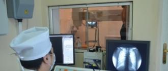
How does it go
X-ray diagnostics are always carried out in a specially equipped, protected room. Before taking a picture, the patient should undress to the waist and remove all metal jewelry (watches, chains and others).

The patient stands in front of a special shield, in which a cassette with a film is installed, is pressed against it closely. The tube from which the x-rays emanate is located approximately two meters away. At the doctor’s signal, you need to inhale and freeze for a couple of seconds.
After the procedure, a person dresses and waits for a medical report.
What do these surveys show
Fluorography with great accuracy reveals tumors, pulmonary tuberculosis, signs of pneumonia and other lesions.
Chest x-ray shows not only these diseases. With the help of x-rays, it is most accurately possible to diagnose tuberculosis, cancer, pneumonia, as well as benign tumors, professional changes. In case of confirmation of the signs of diseases, you should contact the specialized doctor in the clinic to prescribe treatment.
X-ray reveals the pathology of the lymph nodes, some heart diseases, gives information about the condition of the aorta and the inferior vena cava. For a detailed study of disorders in the functioning of the heart and coronary vessels, the patient is sent for echocardiography.
In the X-ray photographs, the bones and joints of the upper body are perfectly visualized.
When planning a pregnancy
Fluorography and x-rays are very well tolerated, usually do not require specialized training, but some contraindications exist.
So, with a planned or confirmed pregnancy, fluorography is not recommended. Received radiation can be dangerous for the embryo. In the first weeks of pregnancy, when the future organs of the child are actively laid, such a test is contraindicated. In the following months, the study is carried out using precautions - shielding the abdomen.

X-ray is a procedure assigned to confirm a preliminary diagnosis, and radiation exposure during radiography, of course, is present. However, if the risk of possible consequences for a woman is assessed higher than for the fetus, the doctor may schedule an examination. Therefore, pregnancy and conception planning cannot be considered absolute contraindications. Moreover, when examining the chest, the danger to the child is many times lower than, for example, with an X-ray or CT scan of the pelvic bones.
If the alternatives are acceptable, then for pregnant women in the first two trimesters an ultrasound scan is recommended, in the 3rd trimester with screening is allowed.
Fluorography and X-rays in childhood
What is better for children: fluorography or x-ray?
Children under 14 years old are not allowed to do fluorography. X-rays are allowed at any age, but they are prescribed it only if the following indications are available:
- cough lasting longer than two weeks;
- suspected pneumonia;
- positive Mantoux reaction.
How many times a year you can do radiography
According to SanPiN 2.6.1.1192-03, everyone should undergo annual fluorography. The only exceptions are children under 14 years of age and pregnant women.
An x-ray is prescribed for suspected diseases localized in the chest area or injuries. There are no restrictions on frequency or dosage. The need for an x-ray is determined by the doctor individually, taking into account the indications and contraindications, as well as taking into account the factors of likely consequences in case of refusal of the procedure.
In case of emergency, x-rays and fluorography can be performed on the same day.
What is the difference between fluorography and x-ray of the lungs
Many people believe that chest x-ray and fluorography are the same thing. Partly they are right. A chest x-ray is the same fluorography that is simply performed on other equipment. The difference is only in the tasks. With fluorography, a routine study is carried out, and with radiography, a clarifying one, since such a diagnosis is more informative. Unnecessarily, they are not carried out simultaneously. If the results of fluorography revealed adverse symptoms, additionally can be assigned:
- x-ray
- cT scan;
- ultrasound procedure;
- endoscopy.
If necessary, fluorography can be replaced by one of the above methods, as is done with children under 14 years of age.
The difference lies in the quality of the pictures. Small lesions or respiratory tract diseases in the early stages of fluorography may not be recorded.
Fluorography is a preventive examination form that is recommended to be taken annually if there are no complaints. An X-ray is prescribed in the presence of symptoms of diseases, revealed pathologies in fluorographic images, and also as a monitoring of the treatment.
What is more harmful x-ray or fluorography
If lung x-rays and fluorography are compared, which method is more harmful? You need to compare the total radiation effect on the body. It all depends not only on the chosen method, but also on the type of equipment. The radiation dose during examination on modern digital equipment is reduced many times, for example:
- when performing fluorography on digital equipment, the exposure rate is only 0.05 mSv;
- if film diagnostics are carried out, then the indicators increase almost ten times (0.3-0.5 mSv).
If we compare x-ray and fluorography on equipment of one class, then when taking pictures, a greater level of exposure comes from the second. But it should be borne in mind that when passing fluorography, only one frame is made. To obtain objective radiographic results, they often take one survey and several aimed shots of the studied area. Thus, the total radiation exposure from x-rays may be higher.

How to check lungs other than fluorography, X-ray
The most common method for studying the lungs, after x-rays and fluorography, is computed tomography. It is also based on x-rays emanating from a tomograph. These rays at various angles reach the internal organs and fall on special hypersensitive sensors. It is they who convert the radiation into an image, which helps doctors get complete information about the patient's condition.
As well as cystography of the urinary system, that is, an x-ray of the bladder, CT of the lungs can be performed using a contrast medium. Indications for this form of research:
- suspected pneumonia;
- benign and malignant tumors;
- primary and secondary metastases;
- pleurisy;
- lymphadenopathy and others.
In some cases, an ultrasound scan is acceptable as an alternative. With the passage of this form of examination, as with duplex scanning of the blood vessels of the liver, it is possible to study the functional state of the vascular bed of the chest area. Simultaneously with ultrasound of the lungs, veins and other vessels of the upper extremities, as well as mammary glands, are often scanned.
Do not forget about endoscopic diagnostic methods. The study of the pleural cavity is carried out under general anesthesia with the help of a tooscope, which penetrates through a small puncture in the chest.